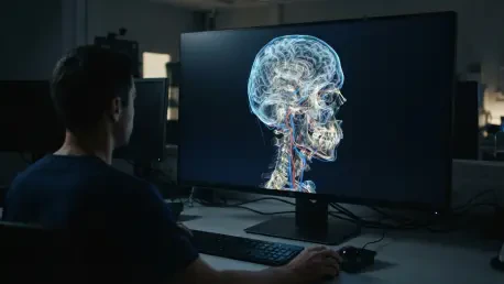

The framework addresses the need for composable and portable APIs in the healthcare domain, allowing for customization across different levels of technical expertise. As medical institutions move away from siloed research environments, the demand for standardized tools that bridge the gap between academic innovation and clinical application has intensified. Conventional computer vision libraries often struggle with the unique nuances of three-dimensional medical scans, such as varying voxel spacing or high-bit-depth modalities. By providing a unified ecosystem, the Medical Open Network for AI establishes a common language for radiologists and data scientists alike. This initiative represents a concerted effort to move beyond fragmented codebases toward a robust, community-driven platform. Consequently, the project focuses on reducing the technical friction associated with handling complex volumetric data. This collaborative spirit ensures that the latest advancements in deep learning are accessible to those working on life-critical imaging tasks.
1. Core Components: Navigating the Project Architecture
The architecture of this project is meticulously organized within the Project-MONAI GitHub organization, where each repository serves a specific function in the deep learning lifecycle. At the heart of the ecosystem lies the primary Python library used for training and testing complex imaging models with medical-specific data transforms. This library extends the capabilities of PyTorch by offering specialized layers and loss functions that are tailored for high-dimensional medical data structures. Simultaneously, the project recognizes the massive bottleneck created by data annotation in the healthcare sector. To resolve this, a dedicated system functions as an intelligent image labeling tool designed to assist research teams with the manual and semi-automated annotation of scans. By integrating with existing viewing software, it allows clinicians to contribute their expertise more efficiently, thereby accelerating the creation of high-quality training datasets that are essential for accurate model development.
Beyond the initial training phase, the ecosystem provides a clear path for translating models into functional clinical utilities. A specialized framework offers a comprehensive set of tools for packaging trained AI models into healthcare-ready applications that follow industry standards. This ensures that a model developed in a laboratory setting can be containerized and verified for deployment within hospital IT infrastructures. For researchers looking to fast-track their progress, a centralized model repository hosts pre-trained weights stored in a standardized bundle format. These models serve as high-performance baselines for various anatomical targets and imaging modalities, saving thousands of compute hours. Complementing these technical tools is a collection of extensive guides and walkthroughs. These educational resources provide the necessary scaffolding for new users to master the API, ensuring that the learning curve remains manageable even as the framework continues to expand its feature set in 2026.
2. Implementation Guide: Installation and Initial Setup
Standardizing the installation process is a priority for the project, ensuring that researchers can establish a functional development environment with minimal friction. The first step involves verifying the local environment to ensure it meets the prerequisite of running Python version 3.10 or a more recent release. This compatibility ensures that the framework can leverage modern Python features and maintain stability across various operating systems. Once the environment is confirmed, the most straightforward method to acquire the stable package is through the Python Package Index using the standard pip command. This approach fetches the latest validated release, providing users with a reliable foundation for their research projects. For those who manage their software dependencies through the Conda ecosystem, the framework is also available via the conda-forge channel. This alternative manager is often preferred in large-scale cluster environments where managing complex dependency trees is critical for system-wide stability.
For teams that require perfectly reproducible environments or operate on high-performance computing clusters, the project offers pre-configured Docker images. Running the official image allows researchers to bypass manual dependency management and gain immediate access to a GPU-optimized environment tailored for heavy computation. After setting up the software, the recommended next step is to explore the practical examples provided in the tutorials repository. These resources illustrate how to integrate domain-specific networks, loss functions, and metrics into a cohesive training pipeline. Mastering these components is essential for effectively handling the complexities of medical imaging data. Finally, users are encouraged to consult the technical manuals and API documentation for their specific version. These documents provide the exact function signatures and parameter definitions required to build custom modules. By following these structured steps, research groups can transition from a fresh installation to active model development.
3. Best Practices: Ensuring Scientific Rigor and Reproducibility
Scientific integrity in the field of medical AI hinges on the ability of independent researchers to replicate findings using identical software configurations and data processing steps. To facilitate this, documenting the specific version of the framework is a mandatory best practice for all published analyses and clinical reports. Users can easily verify their current installation details by querying the package manager, which provides the exact version string necessary for academic disclosure. Relying on the latest unversioned updates can lead to subtle discrepancies in model performance or data transformation outcomes over time. Therefore, maintaining a record of these settings prevents the common pitfall of version drift, which often complicates the peer-review process. When research is conducted within containerized environments like Docker, it is vital to avoid generic tags that may change without notice. Utilizing unique image tags ensures that the computational environment remains frozen in time.
Building on the foundation of version control, the project encourages a modular approach to workflow design that separates data preprocessing from the actual model architecture. This separation allows researchers to swap different components without rewriting entire training scripts, fostering a culture of experimentation and rigorous testing. By utilizing standardized transforms for voxel normalization and spatial orientation, teams can ensure that their data pipelines are consistent across different clinical sites. This level of standardization is particularly important when training models on multi-center datasets where imaging protocols may vary significantly. Furthermore, the framework supports the implementation of validation checks that monitor the consistency of data inputs during the training process. These automated safeguards help identify potential issues early, reducing the risk of training on corrupted or incorrectly formatted medical scans. Adopting these rigorous practices strengthens the credibility of the research.
4. Strategic Evaluation: Advantages, Constraints, and Global Standards
One of the primary strategic advantages of this framework is its permissive licensing structure, which utilizes the Apache-2.0 license to facilitate both academic and commercial utilization. This legal framework provides researchers with the freedom to modify and redistribute the code, encouraging innovation without the restrictive burdens often found in proprietary software. Beyond its legal accessibility, the framework stands out due to its domain-specific design, which is fundamentally different from general-purpose computer vision libraries. Instead of relying on defaults optimized for natural images, it provides specialized transforms and metrics designed for the complexities of medical data, such as 3D rotations and Dice similarity coefficients. Moreover, the library is built to handle the high computational demands of volumetric scans, offering native support for data parallelism across multiple GPUs. This allows researchers to scale their experiments efficiently, reducing training times for even the most complex networks.
The adoption of standardized AI frameworks shifted the trajectory of medical imaging research toward a more collaborative and reproducible future. By integrating modular components and specialized medical transforms, development teams moved beyond the limitations of generic computer vision tools. Practical implementation strategies focused on establishing clear installation protocols and rigorous version control to safeguard scientific integrity. Stakeholders recognized the importance of distinguishing between research-grade tools and regulated medical devices, ensuring that innovation remained aligned with safety requirements. Researchers prioritized the documentation of software versions and the use of containerized environments to ensure long-term stability. This structured approach allowed the community to bridge the gap between initial code development and meaningful clinical insights. Ultimately, the emphasis on standardized citation and versioning fostered a culture where technical innovation and scientific rigor evolved in tandem.